Ottawa Ankle Rules Calculator
This Ottawa ankle rules calculator applies the validated Stiell criteria to decide whether an acute ankle or midfoot injury needs an X-ray. Answer the questions about each injury zone, and the tool tells you whether imaging is recommended by the rule or not indicated by it. It takes less than a minute and follows the exact decision logic published for emergency department use.
Apply the rule: ankle section and foot section
Work through both sections that match where the pain is. Each section has a pain gate followed by its three criteria.
A. Ankle series: malleolar zone
B. Foot series: midfoot zone
How to use this Ottawa ankle rules calculator
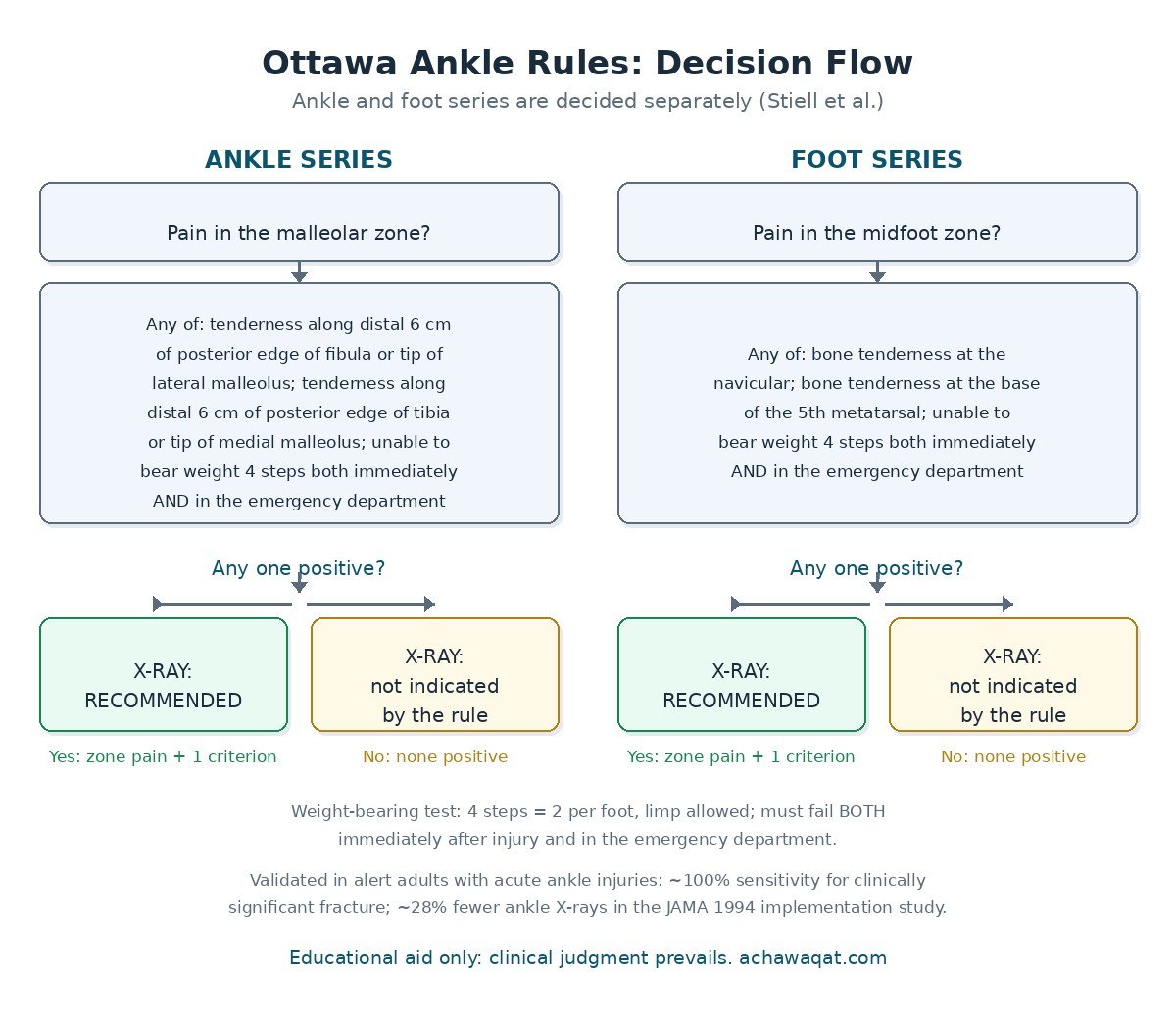
The Ottawa ankle rules are really two rules in one tool, and this calculator keeps them separate on purpose. Section A covers the ankle: it asks first whether there is any pain in the malleolar zone, which is the ring of bone around the ankle joint formed by the ends of the tibia and fibula. Section B covers the foot: it asks first whether there is any pain in the midfoot zone, which is the middle third of the foot between the ankle and the toes. These two pain questions are gates. If the answer is no, that section of the rule simply does not apply, and no X-ray decision can be made for that part of the foot.
If the answer is yes, each section offers its three criteria. You only need one. Bone tenderness at the defined landmarks, or an inability to take four steps both right after the injury and again when examined in the emergency department, each on its own is enough to trigger an X-ray recommendation. Four steps means two steps on each foot, and a limp is allowed, so inability means the person truly cannot do it, not that it hurts. Check every box that applies, then press "Apply the rules" to see the result for the ankle and the foot independently.
The ankle criteria, in plain language
An ankle X-ray series is indicated only when there is pain in the malleolar zone AND at least one of three findings. The first finding is bone tenderness along the distal 6 cm of the posterior edge of the fibula, or at the tip of the lateral malleolus. In practice this means running your thumb up the back edge of the outer ankle bone, from its lowest point to six centimetres above it, and checking for bony pain. The second finding mirrors this on the inner side: bone tenderness along the distal 6 cm of the posterior edge of the tibia, or at the tip of the medial malleolus. The original teaching material stresses palpating the entire 6 cm, not just the tip, and not neglecting the medial side, which examiners are more likely to skip.
The third ankle finding is functional rather than anatomical: the inability to bear weight for four steps both immediately after the injury and in the emergency department. Both moments matter. Some people cannot walk at the scene but manage a few steps once the shock settles, and the rule counts that as able to bear weight. The test is deliberately forgiving, so that only a genuine inability to walk triggers imaging. Together these three criteria make the ankle section of the rule extremely sensitive: in the JAMA implementation study the rule detected every one of the 74 malleolar fractures, a sensitivity of 100 percent.
The foot criteria, in plain language
A foot X-ray series is indicated only when there is pain in the midfoot zone AND at least one of three findings. The first is bone tenderness at the navicular bone, the bony bump on the inner side of the midfoot just ahead of the ankle. The second is bone tenderness at the base of the fifth metatarsal, the bony prominence on the outer edge of the midfoot where the long bone of the little toe begins. These two landmarks are the classic weak points of the midfoot: the navicular takes the force of twisting injuries, and the base of the fifth metatarsal is where avulsion fractures and Jones fractures occur.
The third foot finding is the same weight-bearing test as for the ankle: inability to take four steps both immediately after the injury and in the emergency department. Note that the weight-bearing criterion belongs to both sections, so a patient who cannot walk and has both malleolar and midfoot pain will trigger X-rays of both the ankle and the foot. In the JAMA implementation study the rule detected all 19 midfoot fractures, again a sensitivity of 100 percent. The foot section is sometimes forgotten because the tool is called the ankle rules, which is exactly why this calculator presents both sections side by side.
The two zones: where exactly to look
The zone definitions are part of the rule, not decoration. The malleolar zone covers the distal 6 cm of the tibia and fibula plus the talus, the ankle bone sitting between them. Pain anywhere in this ring, including the front of the ankle, opens the ankle section of the rule, but only tenderness at the specified posterior landmarks counts as a criterion. Pain over the front of the ankle with no posterior tenderness and preserved walking is the classic pattern that the rule says does not need an X-ray, and this is where most of the avoided films come from.
The midfoot zone covers the navicular, cuboid, cuneiform bones, the anterior process of the calcaneum, and the base of the fifth metatarsal. Pain in the toes or the heel does not open the foot section, and tenderness elsewhere in the foot does not satisfy the criteria either. The precision matters because the rule was derived and validated against these exact anatomical definitions; stretching the zones wider would erode the sensitivity that makes the rule safe. When you are unsure whether pain is in the midfoot or the ankle zone, examine both and answer both gates honestly.
How to examine the ankle step by step
Start with the story: how the injury happened, when it happened, and whether the person could walk immediately afterwards. Then inspect both feet and ankles for swelling, bruising, and deformity, comparing sides. Establish the pain zones before you touch anything, because once you start palpating, everything hurts. Ask the person to point to where it hurts most with one finger, and map that onto the malleolar or midfoot zone.
Next, palpate systematically. For the ankle section, feel the posterior edge of the fibula from the tip of the lateral malleolus up 6 cm, then the posterior edge of the tibia from the tip of the medial malleolus up 6 cm. Press on bone, not on the swollen soft tissue in front of it. For the foot section, press the navicular on the inner midfoot and the base of the fifth metatarsal on the outer midfoot. Record bone tenderness only when pressure on the bone itself reproduces sharp pain.
Finally, test weight bearing. Ask the person to take four steps, two on each foot, with or without a limp, and compare with what happened immediately after the injury. If they walked at the scene and can walk now, the criterion is negative even if every step hurts. If they could not walk then and cannot walk now, it is positive. Only then apply the rule: zone pain plus any one positive criterion means imaging, zone pain with everything negative means no imaging is indicated.
Who the rule applies to, and who it does not
The Ottawa ankle rules were derived and validated in alert adults with acute ankle injuries, and that population is where the safety data hold. The original study exclusions, which teaching materials for the rule still carry, include patients under 18, intoxication, painful distracting injuries elsewhere, pregnancy, head injury, and diminished sensation in the legs from a neurological deficit. The logic is straightforward: the rule depends on the patient feeling and reporting pain accurately, so anything that dulls or scrambles that signal makes the rule unreliable.
Clinical judgment should also prevail over the rule in several situations the exclusion lists imply but do not always spell out. Gross swelling that prevents you from feeling the bony landmarks means you cannot apply the tenderness criteria at all. An uncooperative patient, an open wound, obvious deformity, or any sign of neurovascular compromise needs imaging and usually urgent specialist input regardless of what the rule says. Older adults deserve a lower threshold too: they fracture with less force, and several teaching resources advise a liberal use of X-rays in the elderly even when the rule is negative. The rule is a decision aid for the typical adult ankle sprain, not a substitute for examining the patient in front of you.
The evidence behind the Ottawa ankle rules
The rules were derived by Stiell and colleagues and published in the Annals of Emergency Medicine in 1992, from a study that developed the clinical decision rule for radiography in acute ankle injuries. The landmark implementation study followed in JAMA in 1994 (Stiell IG, McKnight RD, Greenberg GH, et al., JAMA 1994;271(11):827-832). It was a nonrandomized controlled trial with before-and-after and concurrent controls, enrolling 2,342 adults with acute ankle injuries across an intervention hospital and a control hospital.
The results are the reason the rule is still taught thirty years later. Ankle radiography fell by a relative 28 percent at the intervention hospital while rising 2 percent at the control hospital, and foot radiography fell 14 percent while rising 13 percent at the control site. Patients discharged without radiography spent less time in the emergency department (80 versus 116 minutes), had lower estimated costs for physician visits and radiography ($62 versus $173), and were just as satisfied with their care (95 versus 96 percent), with identical rates of subsequent radiography (5 percent in both groups). Crucially, sensitivity was 100 percent: the rule caught all 74 malleolar fractures (95 percent confidence interval 0.95 to 1.0) and all 19 midfoot fractures (95 percent confidence interval 0.82 to 1.0). No fractures were missed.
Later research confirmed the finding. A 2003 systematic review in the BMJ (Bachmann LM, Kolb E, Koller MT, et al., BMJ 2003;326:417) pooled the validation studies and found sensitivities between 98.2 and 100 percent, with less than 2 percent of fractures missed when the rule was used. The specificity is low, roughly 42 to 77 percent, which is expected: the rule is designed as a rule-out tool, to make sure no fracture is missed, not to diagnose fractures. A multicentre trial to introduce the rules (BMJ 1995;311:594-597) extended the implementation evidence beyond a single centre. Together this body of work makes the Ottawa ankle rules one of the most validated decision rules in emergency medicine.
Limitations: when clinical judgment wins
The rule only answers one question: is an X-ray indicated to look for a clinically significant fracture, defined in the original work as a bony fragment larger than 3 mm? It says nothing about ligament injuries, which are far more common and are the real diagnosis in most ankle sprains. A negative rule does not mean the ankle is fine; it means no X-ray is needed right now. The standard safety net is written instructions and a recheck in 5 to 7 days if pain and walking ability are not improving, which is what the original teaching material recommends.
The rule also assumes a cooperative, sober adult examined within the acute window after injury. It has not been validated in young children, and several sources advise caution in children under 6. It does not cover injuries of the toes, the heel, or the leg above the malleolar zone, and it does not grade severity. Finally, the weight-bearing test needs the patient to attempt walking, which may be impossible for reasons unrelated to the ankle, such as a knee injury or severe pain elsewhere. In every such case the rule is simply inapplicable, and the clinician decides.
What each result means in practice
When the calculator says an X-ray is recommended, arrange the standard series for that zone: ankle views for a positive ankle section, foot views for a positive foot section, and both when both sections are positive. The recommendation reflects the rule's near-perfect sensitivity, so a positive result should be treated as a genuine indication, not a suggestion. Document which criterion was positive, because the next clinician needs to know why the film was ordered.
When the calculator says an X-ray is not indicated, the evidence says it is safe to forgo imaging, provided the patient fits the rule's population and none of the cautions above apply. Give clear discharge advice: rest, ice, compression, and elevation in the first days, weight bearing as tolerated, and a firm instruction to return or seek recheck in 5 to 7 days if pain is not settling or walking is not improving. A small number of fractures are occult on first presentation, and the follow-up plan is part of the rule's safety, not an afterthought.
Frequently asked questions
Can the Ottawa ankle rules be used in children?
The rules were derived and validated in adults, and the original exclusion lists specify patients under 18. Several later studies support their use in children over 6, with reported sensitivities around 98.5 percent, but the evidence in younger children is limited. In practice, apply the rule cautiously in adolescents, and have a lower threshold for imaging in younger children, whose growth plates and injury patterns differ from adults.
Why does the rule ask about walking both immediately and in the emergency department?
Requiring failure at both moments filters out people who could not walk at the scene from shock or pain but can manage steps once assessed. The rule is deliberately forgiving: a limp is allowed, and four steps means two on each foot. Only a genuine inability at both time points counts as positive, which keeps the criterion sensitive without sending everyone for an X-ray.
Does a negative Ottawa ankle rule mean there is no fracture at all?
No. It means no clinically significant fracture is likely enough to justify an X-ray, where significant was defined in the original work as a bony fragment larger than 3 mm. Tiny avulsion fragments can occasionally be missed, which is why the rule pairs a negative result with written instructions and a recheck in 5 to 7 days if pain or walking ability does not improve. In the JAMA implementation study, no fractures that mattered were missed.
Can I use the rule if the ankle is too swollen to feel the bones?
No. Gross swelling that prevents palpation of the malleolar landmarks is one of the situations where clinical judgment must prevail over the rule. If you cannot feel the posterior edges of the malleoli, you cannot assess the tenderness criteria, so the rule is inapplicable and imaging is the safer course. Re-examine once swelling settles if imaging was deferred.
Do the rules apply to injuries that happened days ago?
The rules were validated in acute injuries presenting to the emergency department, and the weight-bearing criterion specifically asks about the moments right after the injury. For an injury several days old, the "immediately after" part relies on memory and the clinical picture has changed, so the rule is less reliable. A subacute or chronic ankle problem needs a standard clinical assessment rather than this decision rule.
What is the difference between the Ottawa ankle rules and the Ottawa knee rules?
They are sibling decision rules from the same research group, but they cover different injuries and have different criteria. The Ottawa ankle rules decide on ankle and foot X-rays after ankle trauma using malleolar and midfoot tenderness plus weight bearing. The Ottawa knee rules decide on knee X-rays after knee trauma using age, patellar and fibular head tenderness, range of motion, and weight bearing. Do not mix their criteria.
References
- Stiell IG, McKnight RD, Greenberg GH, et al. Implementation of the Ottawa ankle rules. JAMA 1994;271(11):827-832. PubMed PMID 8114236
- Stiell IG, Greenberg GH, McKnight RD, et al. A study to develop clinical decision rules for the use of radiography in acute ankle injuries. Ann Emerg Med 1992;21(4):384-390.
- Bachmann LM, Kolb E, Koller MT, et al. Accuracy of Ottawa ankle rules to exclude fractures of the ankle and mid-foot: systematic review. BMJ 2003;326:417.
- Stiell I, Wells G, Laupacis A, et al. Multicentre trial to introduce the Ottawa ankle rules for use of radiography in acute ankle injuries. BMJ 1995;311(7005):594-597.